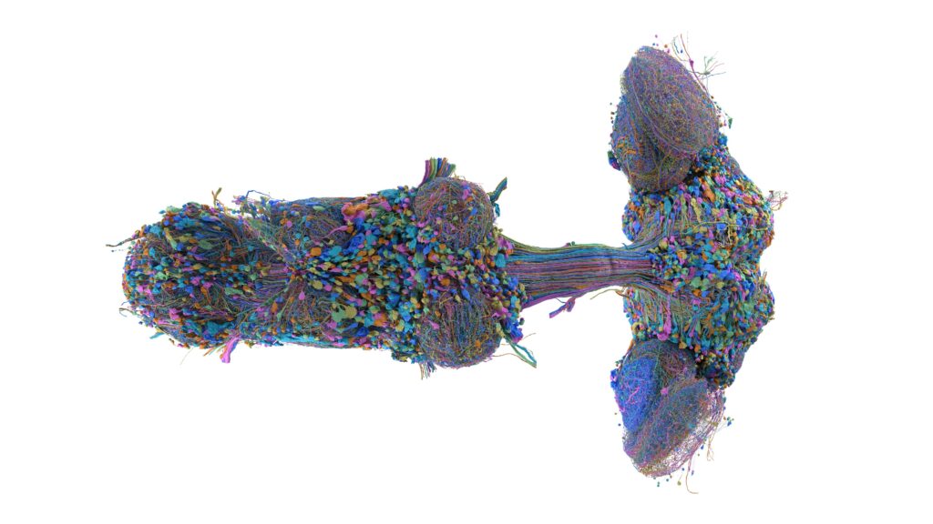
A fruit fly’s entire nervous system now exists as a searchable object: 166,691 neurons and roughly 11,700 cell types, wired together by about 125 million synaptic connections, reconstructed from a single male Drosophila melanogaster by researchers at HHMI’s Janelia Research Campus working with Google Research and collaborators including the Cambridge Connectomics group at the MRC Laboratory of Molecular Biology. In a post published September 3, 2026, Google researchers Michał Januszewski and Viren Jain described the dataset as the first complete wiring diagram of a male fly central nervous system, and the largest brain map by neuron count assembled to date. The accompanying paper in Cell, titled “Sexual dimorphism in the complete connectome of the Drosophila male central nervous system,” lays out the anatomy behind those numbers.
The animal in question weighs less than a milligram. Its brain is smaller than a poppy seed. And every cell in it, plus every wire running down into the equivalent of its spinal cord, has now been traced, named, and made available to anyone with a browser.
What a complete connectome actually contains
The word connectome gets used loosely, so it helps to say what this one is. It is not a functional recording, not a map of activity, not a simulation. It is a reconstruction of physical structure: the shape of each neuron, where its branches go, and which other neurons it touches at synapses — the junctions where one cell passes a chemical signal to the next.
What distinguishes this dataset from earlier fly maps is coverage. Previous reconstructions stopped at the brain. This one includes the brain and the ventral nerve cord, the segmented bundle of tissue that runs through the fly’s thorax and abdomen and plays a role loosely analogous to the spinal cord in vertebrates. That matters because the interesting part of any nervous system is rarely contained in one compartment. A fly seeing a shadow, turning, and beating its wings is running a circuit that starts in the head and ends in muscle, and until both halves were mapped in the same animal, the middle of that circuit was a gap.
The 11,691 cell types in the paper’s count are the organizational layer sitting on top of the raw neurons. Cells are grouped into types by shape and connectivity — neurons that look alike and wire alike get the same label — which turns an unmanageable list of 166,691 individual objects into a vocabulary a researcher can reason with. Assigning those labels was not automated. Human experts at Janelia proofread and annotated the reconstruction, correcting machine errors cell by cell over years.
Machine reconstruction, human correction, and a second sex
The raw material is electron microscopy. The fly’s nervous system was sliced into extremely thin sections and imaged at a resolution fine enough to resolve individual synapses, producing a volume of image data far too large for anyone to trace by hand. Google’s contribution across the decade-long partnership has been the segmentation side: flood-filling networks, a class of AI model that follows the boundary of a single neuron through a stack of images the way water follows a channel, extending a predicted shape voxel by voxel.
The lineage of that approach is visible in earlier milestones. In 2019 the collaboration released an automated reconstruction of a female fly brain. In 2020 came a human-verified reconstruction of roughly half a female brain — about 25,000 neurons and 21 million connections. The current release is a substantial step up in scale, and it arrives with the proofreading already done rather than left as future work.
It also arrives as a counterpart. Because female fly brain and central nervous system maps already exist, the male dataset makes direct comparison possible for the first time, and the Cell authors report exactly that: a large majority of cell types appear in both sexes with matching form — the paper counts 7,205 isomorphic types — alongside 114 types the authors classify as dimorphic, 262 they identify as male-specific, and 69 as female-specific. Read carefully, that ratio is its own finding. Two nervous systems that produce visibly different behaviors are, structurally, mostly the same nervous system with a small set of divergent components.
Why a fly, and what a resource is for
The obvious question is why anyone should care about a poppy-seed brain when the human version holds roughly 86 billion neurons — a scale that, as the Google researchers note, remains far out of reach for this kind of exhaustive reconstruction. The answer is that Drosophila has been a workhorse of genetics and neuroscience for over a century, with a deep toolkit for switching individual neurons on and off. A complete wiring diagram makes that toolkit sharper: a researcher who observes a behavior can now look up which cells could plausibly produce it, and follow the path from sensory input to motor output without guessing at the missing segments.
The dataset is viewable through Neuroglancer, the browser-based tool for navigating large volumetric reconstructions, which means the map is not a figure in a paper but a thing to move around inside. Individual neurons can be pulled out, rotated, and traced to their partners.
Google frames the longer-term hope in medical terms — that understanding how circuits are built and how they fail might eventually inform work on conditions like Alzheimer’s and depression — and it is worth taking that as a stated aspiration rather than a schedule. What exists today is narrower and more concrete: a fully annotated male fly central nervous system, brain and nerve cord together, 166,691 neurons and roughly 125 million connections, described by the teams that built it as the first complete map of its kind, and now open for anyone to search.